备注
转到末尾 下载完整的示例代码。或者通过 Binder 在浏览器中运行此示例。
应用 maskSLIC 与 SLIC#
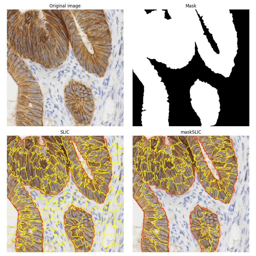
这个例子是关于比较使用普通 SLIC 方法 [1] 和其掩码版本 maskSLIC [2] 获得的分割结果。
为了说明这些分割方法,我们使用了一张经过免疫组织化学(IHC)染色的生物组织图像。在关于如何 免疫组织化学染色中的颜色分离 的示例中,使用了同一张生物医学图像。
maskSLIC 方法是 SLIC 方法在感兴趣区域生成超像素的扩展。maskSLIC 能够克服影响 SLIC 方法的边界问题,特别是在不规则掩码的情况下。

import matplotlib.pyplot as plt

from skimage import data
from skimage import color
from skimage import morphology
from skimage import segmentation

# Input data
img = data.immunohistochemistry()

# Compute a mask
lum = color.rgb2gray(img)
mask = morphology.remove_small_holes(
    morphology.remove_small_objects(lum < 0.7, 500), 500
)

mask = morphology.opening(mask, morphology.disk(3))

# SLIC result
slic = segmentation.slic(img, n_segments=200, start_label=1)

# maskSLIC result
m_slic = segmentation.slic(img, n_segments=100, mask=mask, start_label=1)

# Display result
fig, ax_arr = plt.subplots(2, 2, sharex=True, sharey=True, figsize=(10, 10))
ax1, ax2, ax3, ax4 = ax_arr.ravel()

ax1.imshow(img)
ax1.set_title('Original image')

ax2.imshow(mask, cmap='gray')
ax2.set_title('Mask')

ax3.imshow(segmentation.mark_boundaries(img, slic))
ax3.contour(mask, colors='red', linewidths=1)
ax3.set_title('SLIC')

ax4.imshow(segmentation.mark_boundaries(img, m_slic))
ax4.contour(mask, colors='red', linewidths=1)
ax4.set_title('maskSLIC')

for ax in ax_arr.ravel():
    ax.set_axis_off()

plt.tight_layout()
plt.show()
脚本总运行时间: (0 分钟 0.899 秒)
